Ultrasound microscope identifies cancerous tissue by acoustic profile
3 January 2014
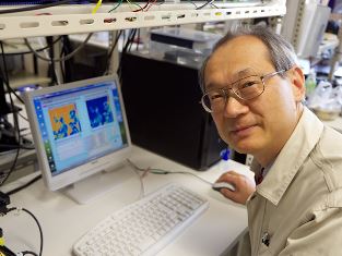
Professor Naohiro Hozumi of Toyohashi University of Technology has developed an ultrasonic microscope to differentiate living tissue and cell specimens for medical purposes.
Ultrasonic microscopes have a wide range of applications including determining the presence of otherwise invisible defects in components used in the automobile, aeronautical, and construction industries.
For medical applications it has advantages over an optical microscope. Whereas an optical microscope is limited to providing only a relative analysis that is based on contrasting shapes of healthy and diseased tissue, the ultrasound technique provides quantitative results based directly on the acoustic properties of tissues.
During surgical operations doctors often stop to inspect tissue taken from a patient’s body for possible remnants signs of disease such as cancer. To do this, pathologists use an optical microscope to examine a slice of tissue taken from the periphery of what should be a healthy area. Now, typical tissues are optically transparent, and must be stained for inspection by an optical microscope. Pathologists can take several hours or possibly several days to evaluate the tissue for the presence of cancerous regions. With an ultrasonic microscope the time can be reduced to a few minutes
“But with my ultrasound microscope, staining is not required because the spectrum of the sound coming back from the tissue changes when the tissue is cancerous, which in turn changes the image,” says Professor Hozumi. “So instead of waiting an hour or more, tissue can be tested almost immediately. Also, because the reflected sound varies depending on the type of cancer, a doctor can interpret the type of disease from the image by comparing it to a reference material.”
“By working quantitatively we can create a database of information,” says Hozumi. “Then, a doctor can use the database to compare the information of a patient’s tissue specimen and readily know whether it is cancerous or not.”

Professor Naohiro Hozumi is developing an
ultrasonic
microscope that can differentiate cancerous cells
This type of procedure requires mounting the removed tissue on a plate for examination under the microscope. Now Hozumi and his colleagues are going a step further by developing an ultrasonic probe. This could be used to directly investigate a patient’s condition immediately after surgery to make sure no cancerous cells remain, and without the need to remove more tissue. The Toyohashi Tech researchers are currently working with microelectromechanical system (MEMS) and semiconductor engineers to develop such devices.