MRI mapping of nerve fibres helps brain surgeons preserve brain function
24 November 2013
A novel technique for interpreting MRI scan data produces images of the brain’s nerve network that can guide neurosurgeons to preserve critical brain functions such as vision, speech and memory.
The technique, called tractography or diffusion tensor imaging (DTI), has been used by University of California San Diego Health System neurosurgeons to guide brain tumour surgery.
Tractography scans, which map the oriented water molecules in nerve fibres, can reveal tiny open paths between the nerve fibres to reach brain tumours. The scans display the neural network in multiple colours. Other current imaging techniques such as computed tomography (CT) and conventional magnetic resonance imaging (MRI) cannot achieve this type of visual display.
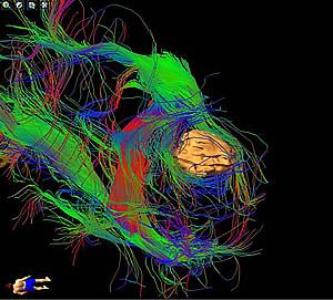
Tractography image showing nerve network around
a brain tumour
“The brain can be mapped by tracking the movement of its water molecules,” said Clark Chen, MD, PhD, neurosurgeon and vice-chairman of neurosurgery at UC San Diego Health System. “Water molecules in brain nerves move in an oriented manner. However, outside the nerves, the molecules move randomly. Neurosurgeons at UC San Diego can use these distinct properties to locate important connections and to guide where surgery should occur or not.”
“There are no margins for error in the brain. Every centimetre of brain tissue contains millions of neural connections so every millimetre counts,” said Chen. “With tractography, we can visualize the most important of these connections to avoid injury. In doing so, we will preserve the quality of life for our patients with brain cancer.”
Anthony Chetti is one of the beneficiaries of tractography-guided brain surgery. Chetti developed a tumour in the region of the brain called the occipital lobe, the portion of the brain responsible for processing visual information. He underwent a complete excision of the brain tumour without any damage to his vision.
“Anytime that you are told that you can potentially lose your vision, you are scared,” said Chetti, a San Diego school teacher. “But when Dr. Chen shared the tractography images with me and showed me how he was going to avoid injury to the connection between my eye and the occipital lobe, I was reassured. When I woke up from surgery, I asked for my glasses immediately and began running systems checks. I could see the clock. I could read the words on a sign. It was immediately evident that there were no problems."
“There is no way of predicting where these fibres would lie except with tractography,” said Chen. “I believe that many brain tumour patients will benefit from tractography-guided surgery. By observing the flow of water molecules, we can visually reconstruct the complex symphony of brain connections. The resulting images are not only accurate but breathtakingly beautiful, giving us a glimpse into the extraordinary human mystery of the brain.”