Multispectral camera highlights cancer remnants for removal during surgery
20 November 2013
Researchers at the Fraunhofer Project Group for Automation in Medicine and Biotechnology (PAMB) have developed a multispectral fluorescence camera system that can make hidden tumour cells visible during surgery.
After removing the main piece of tumour tissue tiny clusters of cancer cells can be left behind as they are difficult to recognize. Tumour margins blend into healthy tissue and are difficult to differentiate. Distributed domains of cancer and pre-malignancies are also difficult to recognize. These pose great challenges even to skilful and experienced surgeons.
Up to now, doctors have depended exclusively upon their trained eyes when excising pieces of tumours. The new camera can help visualize during surgery even the smallest, easy-to-overlook malignant pieces of tumour and thereby support the surgeons during complicated interventions.
Fluorescent dyes
Fluorescent molecules that selectively attach to tumour cells are injected into the patients blood prior to the operation. If the corresponding area is then illuminated with a specific wavelength of light, fluorescence is emitted and the malignant tissue glows green, blue, red, or any other colour, depending on the injected dye, while the healthy tissue appears the same. In this way, the surgeon can see clusters of tumours cells that cannot be recognized by the naked eye.
The new camera can display several fluorescent dyes and the reflectance image simultaneously in real time. This also allows arteries and delicate nerves that must not be injured during an operation to be coloured with a specific dye.
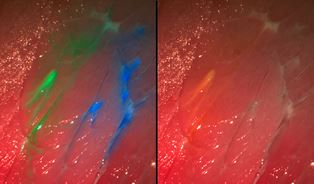
Fluorescence imaging showing tissue dyed in two
colours
In the future the camera will be integrated into various medical imaging systems such as surgical microscopes, endoscopes, etc.
“The visibility of the dye to the camera depends in large part on the selection of the correct set of fluorescence filters. The filter separates the incident excitation wavelengths from the fluorescing wavelengths so that the diseased tissue is also set apart from its surroundings, even at very low light intensities,” says Nikolas Dimitriadis, a scientist at PAMB.
The system requires only one camera and one set of filters for their imaging, which can present up to four dyes at the same time. The team has also developed software that processes the images iand presents it continuously on a monitor during surgery. The information from the fluorescent image is superposed on the normal colour image.
“The operator receives significantly more accurate information. Millimetre-sized tumour remnants or metastases that a surgeon would otherwise possibly overlook are recognizable in detail on the monitor. Patients operated under fluorescent light have improved chances of survival,” says Dr Nikolas Dimitriadis, head of the Biomedical Optics Group at PAMB.
The multispectral fluorescence camera system can be converted to other combinations of dyes. One preparation that is already available to make tumours visible is 5-amino levulinic acid (5-ALA), which is used for glioblastomas, one of the most frequent malignant brain tumours in adults. 5-ALA leads to an accumulation of a red dye in the tumour and can likewise be detected with the camera.
The multispectral fluorescence imaging system should have passed testing for use with humans as soon as next year. The first clinical tests with patients suffering from glioblastomas are planned for 2014.
At MEDICA
PAMB is exhibiting the prototype of the system at the Medica Trade Fair in Düsseldorf in the joint Fraunhofer booth (Halle 10, Booth F05) between 20-23 November 2013.