PET scan tracer highlights areas of pain inside soft tissue
30 October 2013
Researchers at Uppsala University have developed a method to image the areas of damage and pain in soft tissue following injury such as tennis elbow.
It could provide a means to identify true cases of neck whiplash injury in car crashes. This is an area of interest for insurance companies due to the high number of claims for compensation, especially in the UK where it has pushed up the cost of car insurance.
The method involves injecting a radioactive tracer that binds to a signal receptor called NK1 that has been associated with tissue damage. The distribution of radioactive tracer can then be captured as an image using a PET scanner.
Following tissue damage there is an up-regulation of the neuropeptide chemical P and its receptor NK1. This occurs not only in the dorsal horn of the spinal cord, but also in the peripheral painful tissue. This up-regulation is part of an interaction between peripheral nerves, immune cells, and the tissue itself that seems to help guide the body’s own repair process. In chronic tennis elbow, this up-regulation of the substance P-NK1 system lingers on.
This is the first time an up-regulation of NK1 receptors has been visualized by diagnostic imaging in painful tissue in humans. The study clearly reveals an image of elevated levels of NK1 in the painful area compared with the healthy arm.
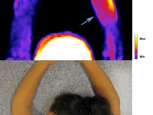
PET image of NK1 receptor radioligand in an
affected arm
Pain from soft tissues (muscles, tendons and ligaments) is still lacking effective methods for localization and diagnosis of underlying pathophysiological mechanisms. This means that diagnosis still depends on clinical examination, which provides no guidance regarding what mechanisms might underlie the pain. Consequently, treatment proceeds purely on an empirical basis. An improved diagnostic method that allows not only diagnosis of localisation of the painful tissue processes, but also can provide guidance regarding what pathophysiological mechanisms are involved, would therefore be highly valuable. The new method is promising, but the costs are still high.
"In the future, we hope to be able to develop less expensive markers that enable us to use the method in everyday clinical practice. We also aim to create markers for other physiological processes that we know are active in chronic soft tissue pain," says researcher Magnus Peterson.
Reference
Peterson M, Svärdsudd K, Appel L, Engler H, Aarnio M, et al. PET-Scan Shows Peripherally Increased Neurokinin 1 Receptor Availability in Chronic Tennis Elbow: Visualizing Neurogenic Inflammation? PLoS ONE 2013 8(10): e75859. doi:10.1371/journal.pone.0075859