Electrosurgical knife instantly detects cancer during surgery
18 July 2013
The iKnife is a surgical knife that cuts flesh using electrical current and analyses the vapour to detect characteristic chemicals of specific cancers using a mass spectrometer.
Invented by Dr Zoltan Takats of Imperial College London, the iKnife can tell surgeons immediately whether the tissue they are cutting is cancerous or not and help them determine which tissue to remove.
The iKnife is based on electrosurgery, a technology invented in the 1920s that is commonly used today. Electrosurgical knives use an electrical current to rapidly heat tissue, cutting through it while minimising blood loss. In doing so, they vaporise the tissue, creating smoke that is normally sucked away by extraction systems.
Dr Takats realised that this smoke would be a rich source of biological information so connected an electrosurgical knife to a mass spectrometer. Different types of cell produce thousands of metabolites in different concentrations, so the profile of chemicals in a biological sample can reveal information about the state of that tissue.
The technology was developed at the Imperial Clinical Phenome Centre, based at St Mary’s Hospital, which was opened last November and is the first of its type in the world. The Centre has a unique collection of technologies for rapid molecular analysis in a hospital setting, aiming to put them at the heart of clinical decision-making. This includes technologies based on mass spectrometry deployed in the operating theatre to give surgeons useful diagnostic information in real-time.
Researchers from Imperial College first used the iKnife to analyse tissue samples collected from 302 surgery patients, recording the characteristics of thousands of cancerous and non-cancerous tissues, including brain, lung, breast, stomach, colon and liver tumours to create a reference library.
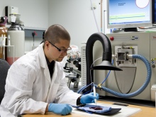
Testing the iKnife in the Clinical Phenome
Centre
at St Mary’s Hospital, London
In 91 tests, the tissue type identified by the iKnife matched the post-operative diagnosis based on traditional methods, but provided the information in less than three seconds, compared to up to half an hour using standard laboratory tests.
In cancers involving solid tumours, removal of the cancer in surgery is generally the best hope for treatment. The surgeon normally takes out the tumour with a margin of healthy tissue. However, it is often impossible to tell by sight which tissue is cancerous. One in five breast cancer patients who have surgery require a second operation to fully remove the cancer. In cases of uncertainty, the removed tissue is sent to a lab for examination while the patient remains under general anaesthetic.
“These results provide compelling evidence that the iKnife can be applied in a wide range of cancer surgery procedures,” Dr Takats said. “It provides a result almost instantly, allowing surgeons to carry out procedures with a level of accuracy that hasn’t been possible before. We believe it has the potential to reduce tumour recurrence rates and enable more patients to survive.”
Although the current study focussed on cancer diagnosis, Dr Takats says the iKnife can identify many other features, such as tissue with an inadequate blood supply, or types of bacteria present in the tissue. He has also carried out experiments using it to distinguish horsemeat from beef.
Professor Jeremy Nicholson, Head of the Department of Surgery and Cancer at Imperial College London, who co-authored the study, said, “The iKnife is one manifestation of several advanced chemical profiling technologies developed in labs our that are contributing to surgical decision-making and real-time diagnostics. These methods are part of a new framework of patient journey optimisation that we are building at Imperial to help doctors diagnose disease, select the best treatments, and monitor individual patients’ progress as part our personalised healthcare plan.”
Lord Ara Darzi, Professor of Surgery at Imperial College London, who also co-authored the study, said: “In cancer surgery, you want to take out as little healthy tissue as possible, but you have to ensure that you remove all of the cancer. There is a real need for technology that can help the surgeon determine which tissue to cut out and which to leave in. This study shows that the iKnife has the potential to do this, and the impact on cancer surgery could be enormous.”
The findings have been published in the journal Science Translational Medicine [1]. The study was funded by the National Institute for Health Research (NIHR) Imperial Biomedical Research Centre, the European Research Council and the Hungarian National Office for Research and Technology.
Reference
1. J. Balog et al. Intraoperative tissue identification using rapid evaporative ionization mass spectrometry. Sci. Transl. Med. 5, 194ra93 (2013).