UCLH hosts endoscopy study day for Pentax Medical’s HD+ and i-scan system
16 August 2012
University College Hospital London NHS Foundation Trust (UCLH) has recently hosted a first ‘hands-on’ gastrointestinal endoscopy study day focusing on the effective use of PENTAX Medical’s HD+ and i-scan imaging system.
The course, attended by consultant endoscopists from around the UK, was presented by UCLH gastroenterology experts. It helped participants to ensure best practice when using i-scan technology and assess the clinical benefits that this technology presents.
UCLH experts, Dr Matthew Banks, Dr Rehan Haidry and Dr Rosa Vega, all lead ‘hands-on’ sessions to help demonstrate the use of i-scan endoscopic optics for both the diagnosis of gastrointestinal mucosal disease and to increase the recognition of early cancer in the upper and lower gastrointestinal tract.
In addition to being able to directly view more advanced endoscopy procedures, such as Endoscopic Mucosal Resection (EMR), delegates also had the opportunity for discussion with peers in a small group environment.
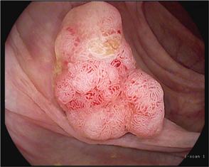
i-scan enhanced image
of Tubulo-villous adenoma
A presentation given by PENTAX Medical product specialists, provided a technical overview of the HD+ and i-scan imaging system. This highlighted key features of the technology which deliver live, clear 1.3 million pixel images and its virtual chromoendoscopy options, for early cancer detection.
Consequently, delegates also participated in detailed tuition on the classification of i-scan Surface Enhanced and Tone Enhanced images using UCLH’s video case study library. Observations of correlations between i-scan images and histology outcomes made during these sessions will contribute to the creation of a new i-scan image classification.
With published evidence now demonstrating that i-scan enhanced images can help to predict early cancer, as presented in a recent paper1 published by the UCLH team, attendees were keen to learn more.
“The study day has been extremely helpful, enabling us to start using the extraordinarily high resolution and virtual chromoendoscopic images produced by i-scan to develop a better way of visually identifying lesions to minimise the use of biopsies and limited histology services in countries such as Zambia,” explained Dr Paul Kelly, Reader in Tropical Gastroenterology, Barts and The London. “I’m concerned that we may be missing early stage cancers that could be identified by using i-scan to its full capacity.”
“I am extremely pleased that our i-scan study day has been very well received by our peers, particularly the opportunity for observation and discussion in small groups during our live cases,” said Dr Matthew Banks, UCLH. “It is great to be able to share our knowledge and experience of the clinical benefits presented by HD+ and i-scan imaging. We also appreciate the feedback from attendees to ensure that future study days will even better meet the educational needs of our delegates.”