Ultrasound identifies inflammatory regions in tendinitis
8 March 2011
Prof Carlo Faletti of the Maria Adelaide Hospital, Turin has validated the benefits of using ultrasound over traditional technologies, like MRI (the reference technology), for the diagnosis of the inflammatory disease tendinitis.
The disease is caused primarily by sporting accidents, overuse and degenerative pathologies.
Prof Faletti, Director of the Diagnostic Imaging Department at the hospital, supported by ultrasound system developer Aloka, studied the advantages of conducting assessments using ultrasound combined with e-flow (an imaging function unique to Aloka). The findings demonstrate that ultrasound can enable the radiologist to view a patient’s injuries in real-time — which is a big advantage when trying to identify the precise location of acute pain, and thus its true cause.
Although the findings showed MRI and ultrasound produced very similar diagnostic information, using e-flow produced greater sensitivity and far greater image detail. During the acute phase Prof. Faletti was also able to observe micro-angiogenesis (the growth and repair of new blood vessels) taking place, which provides important clinical outcomes on a tendon’s insertion (eg supraspinatus tendon).
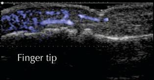
Ultrasound image of inflammation in a finger
tip
A major part of Prof. Faletti’s work revolves around working with prominent sporting stars and a leading Italian Serie A football club, and he has now taken to adopting a ‘fused’ approach using both MRI and ultrasound; as with e-flow he can pin-point with great accuracy any bleeding in fascial regions (connective tissue that surrounds muscles).
Additionally, Prof. Faletti believes in the future, especially with new models like the Aloka ProSound F75, we will be able to view even greater detail to detect new pathologies — such as 3D reconstruction of the tissue structure and lesions.
Prof Faletti, commented: “High resolution ultrasound, in combination with Contrast Enhanced Ultrasound (CEUS) and e-flow technologies is a highly sensitive diagnostic method for the assessment of patellar and achilles insertiional tendinopathies. Having the ability to ask a patient where they feel pain, whilst watching in real-time imaging has allowed me to get a better understanding of the clinical situation — and I would advice all clinicians to consider working with ultrasound when evaluating conditions like these.”