Maths and video game graphics help develop live MRI of beating heart
2 Sept 2010
Magnetic resonance imaging taking just one fiftieth of a second have been recorded by researchers at the Max Planck Institute for Biophysical Chemistry, Göttingen.
With this breakthrough, the dynamics of organs and joints can be filmed 'live' for the first time, including the beating heart, the bending knee, and movements of the eye and jaw.
The new MRI method promises to add important information about diseases of the joints and the heart. In many cases MRI examinations may also become easier and more comfortable for patients.
The recording of cross-sectional images of the body by magnetic resonance imaging (MRI) required several minutes until well into the 1980s, but now only takes a matter of seconds. This was enabled by the FLASH (fast low angle shot) method developed by Göttingen scientists Jens Frahm and Axel Haase at the Max Planck Institute for Biophysical Chemistry.
FLASH revolutionised MRI and was largely responsible for its establishment as a most important modality in diagnostic imaging. MRI is completely painless and, moreover, extremely safe. Because the technique works with magnetic fields and radio waves, patients are not subjected to any radiation exposure, as is the case with X-rays.
At present, however, the procedure is still too slow for the examination of rapidly moving organs and joints. For example, to trace the movement of the heart, the measurements must be synchronised with the electrocardiogram (ECG) while the patient holds the breath. Afterwards, the data from different heart beats have to be combined into a film.
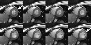
Real-time MRI of the heart with a measurement
time of 33 milliseconds per image and 30 images per second. The
spatial resolution is 1.5 millimetres in the image plane (section
thickness 8 millimetres). The eight successive images show the
movement of the heart muscle of a healthy subject for a period of
0.264 seconds during a single heartbeat. The images range from the
systolic phase (arrow, top left: contraction of the heart muscle) to
the diastolic phase (arrow, bottom right: relaxation and expansion).
The bright signal in the heart chambers is the blood.
Future prospect: extended diagnostics for diseases
The researchers working with Jens Frahm, Head of the non-profit Biomedizinische NMR Forschungs Gmb”, succeeded in further accelerating the image acquisition process. The new MRI method developed by Jens Frahm, Martin Uecker and Shuo Zhang reduces the image acquisition time to one fiftieth of a second (20 milliseconds), making it possible to obtain live recordings of moving joints and organs at so far inaccessible temporal resolution and without artefacts.
Filming the dynamics of the jaw during opening and closing of the mouth is just as easy as filming the movements involved in speech production or the rapid beating of the heart.
“A real-time film of the heart enables us to directly monitor the pumping of the heart muscle and the resulting blood flow — heartbeat by heartbeat and without the patient having to hold the breath,” explains Frahm.
The scientists believe that the new method could help to improve the diagnosis of conditions such as coronary heart disease and myocardial insufficiency. Another application involves minimally invasive interventions which, thanks to this discovery, could be carried out in future using MRI instead of X-rays.
“However, as it was the case with FLASH, we must first learn how to use the real-time MRI possibilities for medical purposes,” says Frahm. “New challenges therefore also arise for doctors. The technical progress will have to be ‘translated’ into clinical protocols that provide optimum responses to the relevant medical questions.”
Less is more: acceleration through better image reconstruction
To achieve the breakthrough to MRI measurement times that only take very small fractions of a second, several developments had to be successfully combined with each other.
Whilst still relying on the FLASH technique, the scientists used a radial encoding of the spatial information which renders the images insensitive to movements. Mathematics was then required to further reduce the acquisition times.
“Considerably fewer data are recorded than are usually necessary for the calculation of an image. We developed a new mathematical reconstruction technique which enables us to calculate a meaningful image from data which are, in fact, incomplete,” explains Frahm.
In the most extreme case it is possible to calculate an image of comparative quality out of just 5% of the data required for a normal image — which corresponds to a reduction of the measurement time by a factor of 20. As a result, the Göttingen scientists have accelerated MRI from the mid 1980s by a factor of 10000.
Although these fast MRI measurements can be easily implemented on today’s MRI devices, something of a bottleneck exists when it comes to the availability of sufficiently powerful computers for image reconstruction.
Physicist Martin Uecker explains: “The computational effort required is gigantic. For example, if we examine the heart for only a minute in real time, between 2000 and 3000 images arise from a data volume of two gigabytes.”
Uecker consequently designed the mathematical process in such a way that it is divided into steps that can be calculated in parallel. These complex calculations are carried out using fast graphical processing units that were originally developed for computer games and three-dimensional visualization.
“Our computer system requires about 30 minutes at present to process one minute’s worth of film,” says Uecker. Therefore, it will take a while until MRI systems are equipped with computers that will enable the immediate calculation and live presentation of the images during the scan. In order to minimise the time their innovation will take to reach practical application, the Göttingen researchers are working in close cooperation with Siemens Healthcare.
References
1. Martin Uecker, Shuo Zhang, Dirk Voit, Alexander Karaus, Klaus-Dietmar
Merboldt, Jens Frahm. Real-time MRI at a resolution of 20 ms.
NMR in Biomedicine 23. doi:10.1002/nbm.1585 (Online 27 August
2010)
onlinelibrary.wiley.com/journal/10.1002/%28ISSN%291099-1492/earlyview
2. Shuo Zhang, Martin Uecker, Dirk Voit, Klaus-Dietmar Merboldt,
Jens Frahm. Real-time cardiovascular magnetic resonance at high
temporal resolution: radial FLASH with nonlinear inverse
reconstruction. Journal of Cardiovascular Magnetic Resonance
12, 39 (2010). doi:10.1186/1532-429X-12-39
www.jcmr-online.com/content/12/1/39