|
|
|
| Diagnostic imaging, neurology | |
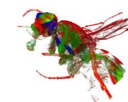
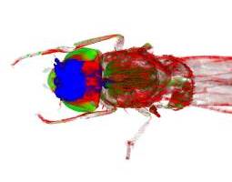
Fruit fly imaging aids research into Alzheimer's18 September 2007 Scientists at the UK Medical Research Council (MRC) have developed optical imaging technology that can generate 3D internal images of the fruit fly for the first time. Using an imaging technique called optical projection tomography (OPT), which was originally developed at the MRC Human Genetics Unit, they have generated startling 3D images of the inside of a fruit fly. Due to the similarity between human and fly genes, the technique could help to speed up genetic research into Alzheimer’s and other human diseases that affect brain cells.
Dr Mary O’Connell of the MRC Human Genetics Unit who led the research explained: ‘‘Neurodegeneration, the gradual loss of function of brain cells that occurs in Alzheimer’s, Parkinson’s and motor neurone diseases, isn’t a strictly human phenomenon. Insects are affected by it too. In the autumn, bees and wasps often develop erratic behaviour before they die." "It’s already known that defects in the equivalent fly genes involved in human brain diseases cause brain cells in fruit flies to lose function as they age,’’ Dr O’Connell continued. Because the fruit fly (Drosophila melanogaster) and humans share many genes with similar functions, the fly is widely used by genetic researchers to study how genes influence human disease. OPT could help researchers to look at how the fly brain changes in
response to alterations in the normal activity of a specific gene without
the risk of damaging tissue through dissection. MRC PhD student Leeanne McGurk who captured many of the OPT images explained why the technique works: ‘‘The dark colour of the fly exoskeleton prevents us from seeing inside it using a standard light microscope. In the past this has meant scientists have had to tease apart fruit fly tissues by hand — a laborious process. Now, we have got over the problem by bleaching the fly exoskeleton. When the fruit fly becomes colourless it is possible to use imaging techniques not only to view its internal organs but to generate 2D and 3D images of the entire fly. ’’ Using OPT images in this way will allow scientists to visualise where and how the products of selected genes are present in the fly. These patterns of gene expression, will help to identify genes that control parts of the central nervous system and so provide detailed information about the human brain. Bleaching of the exoskeleton to clear away the colour also allows images to be generated using other microscopic techniques that depend on penetration of light. Dr O’Connell concluded: ‘‘This research is not simply limited to the study of conditions like Alzheimer’s but can also be used to study fly anatomy. The shape and size of organs can be affected by diseases like diabetes so imaging may yield clues to further our understanding of other conditions too.’’ Reference 1. McGurk L, Morrison H, Keegan LP, Sharpe J, O’Connell MA (2007) Three-Dimensional Imaging of Drosophila melanogaster. PLoS ONE 2(9):e834. doi:10.1371/journal.pone.0000834 The paper is available at: www.plosone.org/doi/pone.0000834
|