| Diagnostic imaging |
|
|
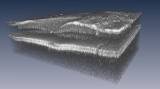
Rapid laser scanning produces high resolution 3-D images of living retina9 May 2007 Researchers at the Massachusetts Institute of Technology (MIT) have produced a new laser that can scan the living eye at record speeds and produce a 3-D image of the retina in less than a second. The retinal imaging is performed with an emerging method called optical coherence tomography (OCT), which uses light to obtain high-resolution images of the eye, even for structures such as the retina that lie beneath the surface. The retina converts light to electrical signals that travel to the brain.
Conventional OCT imaging typically yields a series of two-dimensional cross sectional images of the retina, which can be combined to form a 3-D image of its volume. Even more helpful for diagnosing disease would be to obtain very-high-resolution three-dimensional views of the eye. Limited imaging speeds and involuntary eye motion (such as blinking) make it difficult to perform 3-D imaging of the retinal volume. In ophthalmology applications, OCT systems work by scanning light back and forth across the eye, tracing thin, micrometer-scale lines that row by row build up high-resolution images. Commercial systems scan the eye at rates ranging from several hundred to several thousand lines per second. A typical patient can only keep the eye still for about one second, limiting the amount of three-dimensional data that can be acquired. Robert Huber (now at the Ludwig Maximilians University in Germany) and colleagues at the MIT have reported retinal scans at record speeds of up to 236,000 lines per second, a factor of 10 improvement over current OCT technology. With their technique, which uses a frequency-tunable laser to achieve fast scan speeds, they obtained a 3-D retinal image consisting of 512x512x400 volume elements of data in a human subject in just 0.87 seconds. Future clinical studies, as well as further development, may someday enable ophthalmologists to routinely obtain three-dimensional "OCT snapshots" of the eye, containing comprehensive volumetric information about the microstructure of the retina. Such snapshots could potentially improve diagnoses of retinal diseases such as diabetic retinopathy, glaucoma and age-related macular degeneration. This research will be presented at CLEO/QELS in Baltimore, on 10 May. Meeting Paper: CThAA5, "Fourier Domain Mode Locking (FDML) in the Non-Zero Dispersion Regime: A Laser for Ultrahigh-Speed Retinal OCT Imaging at 236kHz Line Rate," Huber et al., Thursday, May 10, 3:45 p.m. - 4:15 p.m.
|