| Diagnostic imaging | |||
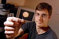
New cellular sensing tool aids cystic fibrosis research31 March 2006 Atlanta, USA. A new microelectrode that combines an atomic force microscope with a biosensing layer has been used to investigate the cellular energy transport chemical adenosine triphosphate (ATP) and its role in cystic fibrosis. The ATP study is the first application of the novel sensing system developed by a research team led by Christine Kranz a senior research scientist at Georgia Institute of Technology.
This patented technology adds recessed micro- and nano-electrodes to the
tip of an atomic force microscope (AFM), creating a single tool that can
simultaneously monitor topography along with electrochemical activity at the
cell surface. “Conventional AFM can image surfaces, but usually provides limited chemical information,” he explained. “And though scanning electrochemical microscopy (SECM), another probing technique, provides laterally resolved electrochemical data, it has limited spatial resolution. By combining AFM and SECM functionality into a single scanning probe, our tool provides researchers with a more holistic view of activities at the cell surface.” In addition to Mizaikoff and Kranz, the team also includes post-doctoral scholar Jean-Francois Masson and graduate student Justyna Wiedemair. In the ATP study, which is sponsored by the National Institutes of Health and done in collaboration with Douglas Eaton at Emory University’s School of Physiology, the Georgia Tech team used the multi-scanning biosensors to study ATP release at the surface of live epithelial cells (cells that cover most glands and organs in the body). ATP, a chemical involved in energy transport, is of interest to medical researchers because elevated levels have been linked with cystic fibrosis, a disease that affects one out of every 2,500 people in the United States. Using epithelial cell cultures from Emory, the Georgia Tech researchers have demonstrated that their multi-functional biosensors work at the live-cell surface during in vitro studies. “Before you can identify what triggers the ATP release, we must be able to quantitatively measure the released species at the cell surface,” Mizaikoff said, noting that many pathological events involve the disruption of chemical communication and molecular signalling between cells, especially in the nervous system, lungs and kidneys. Improved understanding of cellular communication can lead to new strategies for treating diseases, Mizaikoff added: “Being able to operate sensors in an electrochemical imaging mode at the micro- and nanoscale is an exciting opportunity for complementing optical imaging techniques. There are many clinical research problems that these biosensors can help with.”
|