| Diagnostic imaging | |
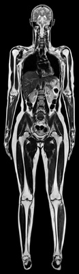
Total imaging matrix reveals new diagnostic possibilities for MRI16 September 2005 Total imaging matrix (Tim) is a new method of magnetic resonance tomography introduced by Siemens Medical Solutions in 2004. It allows whole-body imaging in a single examination with excellent image quality. Nearly 600 Tim systems have now been installed worldwide and the first clinical experiences confirm the broad performance spectrum of this technology.
Tim technology greatly enhances cardiac examinations. The use of a 32 channel coil and new acquisition methods such as AutoViability and Cine Late Enhancement allow for a display of the infarcted area during heart contraction, facilitate the examination as such, and improve the diagnosis of cardiovascular diseases with MR. Not only is it possible to obtain higher resolution, the workflow shows considerable improvement as well: today, a full cardiac examination of an infarcted patient can be completed within 15-30 minutes. The in the US already patented technique for precisely displaying infarcts with Delayed Enhancement, developed together with Raymond Kim, MD of the Cardiovascular Magnetic Resonance Center at Duke University, Raleigh Durham, North Carolina, USA, has opened a new field of indications, as shown by several international cooperation partners who are now able to diagnose and differentiate various types of cardiomyopathy with CMR. This examination helps determine the risks of an untreated patient to die from sudden cardiac death. These statements are confirmed by the work of Professor Dudley Pennell of the Royal Brompton Hospital in London. This information can be an important decision-making tool for implanting a pacemaker.
Pennell was also able to change the invasive therapeutic monitoring of thalassemia patients — by biopsying the liver — to a non-invasive method using MR. Thalassemia, a special form of cardiomyopathy, is a genetic disease prevalent in Mediterranean and Asian countries. Untreated, the disease is fatal. However, treatment of the disease leads to iron deposits in the heart and liver which lead to heart failure and sudden cardiac death. To prevent this outcome, the iron load in the heart has to be checked carefully. Until now, only biopsies provided the necessary results. But these were not taken from the heart, but rather from the liver. However, since the heart is the decisive factor in this disease, the morbidity rate for patients receiving treatment remained very high. For the first time, it is now possible to determine the iron contents of the heart in five minutes with Magnetic Resonance Tomography and Tim. Since it is now possible to control these effects, the morbidity rate has decreased considerably.
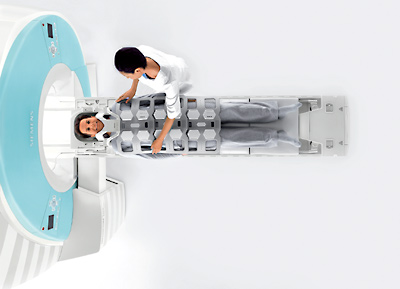
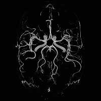
In addition, an examination with Tim allows for a multi territory assessment through out of cardiovascular diseases throughout the entire body in a single exam without patient repositioning, for example arteriosclerosis and diabetes do not affect the heart alone, but other organs and vessel systems as well. For diabetes, the kidneys in connection with the heart and the entire vascular system can be examined in a single exam in high resolution. The physician is able to diagnose vessel changes early and take therapeutic measures before the heart muscle is damaged. Although Tim examines patients who are as tall as 6 feet 9 inches, the technology also offers special advantages when dealing with children: using measurement techniques such as GRAPPA and T-SENSE combined with the 32 RF channels of Tim, the fast heart rates of children as well as the smallest anatomical structures are no longer a challenge. For this reason, Tim systems are frequently used in specialty hospitals and departments for paediatric cardiology to diagnose and plan operations for congenital heart defects. The system allows for a four-dimensional display of the heart and vessels at an improved spatial resolution of below one millimetre at an even shorter acquisition time. In addition to the Magnetom Avanto, other Siemens MR systems are now equipped with the Tim technology as well. Recently, the first two 3 Tesla MR systems, the Magnetom Trio, were equipped with Tim and put into operation in the US and Germany. The Magnetom Espree, the shortest 1.5 Tesla system worldwide with a very open bore diameter of 70 cm and only 1.25 m length was introduced last summer. In combination with Tim, the system examines the effects of overweight and obesity on the heart, vessels and other organs with a higher image quality than usually obtained with "open" systems. Overweight people run three times the risk of dying from a heart attack. Another effect of the systems’ short magnet length is that the patient finds himself in a larger space. This provides the physician with better access to the patient, a fact that is of decisive importance during cardiac examinations using pharmacological stress. |